The other groove is the anterior interventricular sulcus situated on the sternocostal surface of the heart close to its left margin. Which heart groove travels between the atria and the ventricles a anterior interventricular sulcus b.
![]()
Posterior Interventricular Artery Anatomy And Supply Kenhub
Posterior interventricular sulcus Shows the surface boundaries between the two ventricles on the posterior surface of the heart.
. These extend into the auricle. Locate the right and left ventricle and right and left atrium. Is another name for the coronary sulcus.
Located between the left and right ventricles are two additional sulci that are not as deep as the coronary sulcus. Called also interventricular groove. Left anterior descending artery bifurcation.
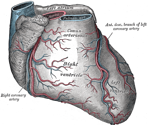
Starting from the apex of the heart and running parallel with the anterior interventricular artery the great cardiac vein travels up along the anterior interventricular sulcus towards the base of the left atriums auricle. Left and right atrioventricular grooves or coronary sulcus 2. The phrenic nerve may lie close to the lateral.
Posterior interventricular sulcus c. Coronary sulcus both a and b e. Right atrium The vessels that carry oxygen poor blood deoxygenated to the heart empty into the.
Is a groove between the ventricles on the back of the heart. Anterior and posterior interventricular sulcus C Coronary sulcus. Anterior interventricular sulcus 3.
The other groove is the anterior interventricular sulcus situated on the sternocostal surface of the heart close to its left margin. The anterior interventricular sulcus is situated on the sternocostal surface of the heart close to its left margin. Where is the base of the heart.
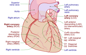
In it runs the posterior interventricular artery and middle cardiac vein. Download Unionpedia on your Android device. When it reaches the apex it anastomoses with the anterior interventricular artery a branch of the left coronary artery.
Notice the anterior interventricular sulcus posterior interventricular sulcus Posterior 4. The posterior interventricular sulcus or posterior longitudinal sulcus is one of the two grooves that separates the ventricles of the heart and is on the diaphragmatic surface of the heart near the right margin. The anterior interventricular sulcus or anterior longitudinal sulcus is one of two grooves that separates the ventricles of the heart the other being the posterior interventricular sulcus.
What is the function of the posterior interventricular artery. A b and c 3. Posterior interventricular sulcus and Anterior interventricular sulcus See more Heart.
Parallels the anterior interventricular artery and drains the areas supplied by this vessel heart block interruption in the normal conduction pathway heart bulge. The adult heart structure that marks the location of an opening between the two atria in the fetal heart is called. Free Faster access than browser.
After crossing the posterior interventricular sulcus it directly continues as the great cardiac vein GCV that runs around the anterior and lateral portions of the mitral annulus GCV was divided into three segments proximal middle and distal by lateral vein LV and posterior-lateral vein PLV the distal great cardiac vein DGCV was. What vessels are situated in sulcus Interventricularis posterior. The posterior interventricular sulcus.
Distinguish the four chambers of the heart. The other groove is the anterior interventricular sulcus situated on the sternocostal surface of the heart close to its left margin. Either of the anterior and posterior grooves on the surface of the heart that lie over the interventricular septum and join at the apex.
Lay the heart on the dissecting tray anterior surface up. Contains the great cardiac vein and coronary sinus. The anterior interventricular sulcus or anterior longitudinal sulcus is one of two grooves that separates the ventricles of the heart the other being the posterior interventricular sulcus.
Asked Sep 27 2015 in Anatomy Physiology by Dylan. What structures does the posterior interventricular artery supply. Once the vein reaches the left margin of the heart it circumvents to the posterior side and eventually converges with the oblique vein of the left.
Medical Definition of interventricular sulcus. Base posterior surface - formed by the atria mainly the left atrium B. The other groove is the anterior interventricular sulcus situated on the sternocostal surface of the heart close to its left margin.
Vessel that follows the interventricular sulcus on the anterior surface of the heart and flows along the coronary sulcus into the coronary sinus on the posterior surface. In certain patients the left anterior descending artery borders the apex and continues through the posterior interventricular sulcus to its middle third irrigating much of the lower wall. In these cases the posterior descending artery usually has a small diameter and is short.
The anterior interventricular sulcus or anterior longitudinal sulcus is one of two grooves that separates the ventricles of the heart the other being the posterior interventricular sulcus. Coronary sulcus and posterior interventricular sulcus E. The anterior interventricular sulcus or anterior longitudinal sulcus is one of two grooves that separates the ventricles of the heart the other being the posterior interventricular sulcus.
The posterior interventricular artery descends in the interventricular sulcus towards the apex of the heart. The crux of the heart is the meeting point of the interatrial and interventricular septa of the cardiac chambers. Clinical Significance The coronary arteries can vary with respect to their origin number and course and many variants have been described in medical literature.
The anterior interventricular sulcus is situated on the sternocostal surface of. The anterior interventricular sulcus is visible on the anterior surface of the heart whereas the posterior interventricular sulcus is visible on the posterior surface of the heart. The anterior interventricular sulcus is situated on the sternocostal surface.
Anterior interventricular sulcus Shows the separation of the two ventricles on the anterior surface of the heart. It drains a significant portion of the LV anterior wall and the interventricular septum begins at the cardiac apex and ascends toward the base of the heart in the anterior interventricular sulcus parallel to the left anterior descending coronary artery. The epicardium and the 4.
The anterior interventricular sulcus is situated on the sternocostal surface of the heart close to its left margin. The anterior interventricular vein is the largest and most consistent of the cardiac veins. The anterior interventricular sulcus or anterior longitudinal sulcus is one of two grooves that separates the ventricles of the heart the other being the posterior interventricular sulcus.
The anterior interventricular sulcus or anterior longitudinal sulcus is one of two grooves that separates the ventricles of the heart the other being the posterior interventricular sulcus. Is a valve in the interventricular septum that closes at birth. The posterior interventricular sulcus or posterior longitudinal sulcus is one of the two grooves that separates the ventricles of the heart and is on the diaphragmatic surface of the heart near the right margin.
The posterior interventricular artery supplies blood to the hearts posterior or bottom portion. The posterior interventricular sulcus or posterior longitudinal sulcus is one of the two grooves that separates the ventricles of the heart and is on the diaphragmatic surface of the heart near the right margin. Notice the Posterior Dorsal View.
![]()
Posterior Interventricular Artery Anatomy And Supply Kenhub

Posterior Interventricular Artery An Overview Sciencedirect Topics

A P Ii Lab Lab Exam Heart Anatomy Flashcards Quizlet

Posterior Interventricular Artery An Overview Sciencedirect Topics

Veins Sulcus Of Heart Anterior View Diagram Quizlet
Anterior Interventricular Sulcus Wikipedia
Posterior Interventricular Artery Wikipedia

Posterior Heart Surface Circulatory System Heart Structure Heart Anatomy
0 comments
Post a Comment